
MS Progression May Be Tied to Workings of Immune Complement System in Brain Lesions
Written by |
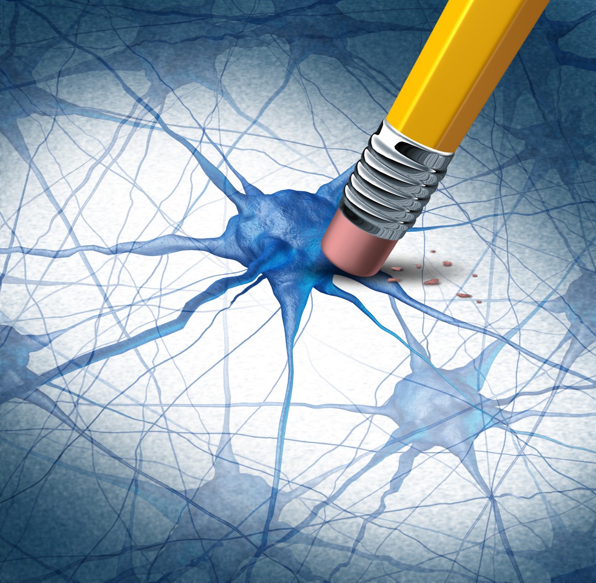
The complement system, a part of our non-adaptable (innate) immune defenses, is activated in lesions inside the brain’s gray matter and may well contribute to the relentless progression of multiple sclerosis (MS), researchers report. The findings offer new insights into mechanisms driving the development of this disease — particularly its primary progressive forms.
The complement system is composed of an array of proteins that, by triggering a cascade of molecular signals, enhances (complements) the activity of other parts of the immune system, including antibodies and cells that clear microbes and cell debris.
High levels of complement system factors are found in blood and cerebrospinal fluid of MS patients, suggesting that the system is actively contributing to disease. But precisely how it contributes is not at all clear. Earlier studies have shown that — in contrast to brain lesions in the myelinated white matter of the brain — increased numbers of lesions in the gray matter predicts disease course.
During brain development, the complement system is needed for synaptic pruning, a process whereby excessive brain connections are eliminated, making researchers suspect that the same mechanisms might be involved in neurodegeneration in MS. A few studies have confirmed this by showing the presence of certain activated complement factors, along with a lack of molecules controlling them, in brain areas marked by nerve cell death. So far, however, no studies have done more extensive screening of complement factors in the brains of MS patients.
Researchers at Swansea University School of Medicine in the U.K. analyzed a large set of complement factors, receptors, and regulatory molecules in the brains of 22 deceased MS patients and 23 control individuals, of which 15 did not have neurological disease and eight had other types of inflammatory disease.
The study, “Complement is activated in progressive multiple sclerosis cortical grey matter lesions,“ published in the Journal of Neuroinflammation, found high levels of activated complement in gray matter lesions. Such lesions could be detected both in the surface layer of the brain, called the cortex, and in deeper brain regions.
Researchers noted that microglial cells — a brain cell type involved in immune processes — carried complement receptors on their surfaces and were present in high numbers in the lesions. In lesioned areas with high levels of complement factors, the nerve cells also looked different, showing signs of stress and neurodegeneration. The team found that the extent of nerve cell loss in a brain area was linked to the numbers of cells with complement factors in that area.
“Complement is activated in the MS cortical grey matter lesions in areas of elevated numbers of complement receptor-positive microglia and suggests that complement over-activation may contribute to the worsening pathology that underlies the irreversible progression of MS,” the research team concluded.


Leave a comment
Fill in the required fields to post. Your email address will not be published.