New MRI Method Has Potential to Map MS Progression and Guide Treatment
Written by |

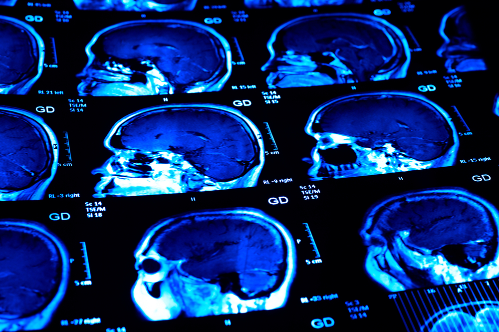
Researchers working with magnetic resonance imaging (MRI) are often faced with a problem: an average MRI brain scan produces a considerable amount of images (around 600 megabytes), but half carry distortions that make them unreadable. These “phase images,” as they are known, are usually discarded and their insights lost. Now, the work of researchers like Hongfu Sun in the field of quantitative susceptibility mapping (QSM) is revolutionizing the MRI field, and information within “phase images” is no longer a mystery.
This means that for diseases such as multiple sclerosis (MS), researchers could soon be much better equipped to track biomarkers in the brain that correspond with the severity of MS symptoms.

Postdoctoral scholar Hongfu Sun works in multiple sclerosis assessment. (Photo by Riley Brandt, University of Calgary)
Dr. Sun, who received a T. Chen Fong Postdoctoral Fellowship for his innovative MRI methods targeting MS assessment, is now developing his research in Dr. Bruce Pike’s lab, part of The Hotchkiss Brain Institute (HBI) at the Cumming School of Medicine, Canada.
“Hongfu has already established himself as a creative thinker during his PhD and his innovative contributions have received significant attention,” Dr. Pike said in a university press article by Pamela Hyde. “He is a curious and driven young scientist with enormous potential and I am delighted to have him join my team.”
MRI scans are very useful in diagnosing MS, because they identifies lesions where myelin has been destroyed. But that is all they can do.
“If you just count how many lesions there are, it doesn’t correlate with how bad the disease is. For many years people have struggled to find a biomarker that could correlate with the disease,” Dr. Sun said.
During his PhD, Dr. Sun discovered that iron accumulation in the brain, specifically in the brain’s deep grey matter, was associated with worsening MS symptoms. These findings are the foundation of his postdoctoral research, where he will use the MRI methods he developed to measure myelin sheaths, located in the brain’s white matter.
“Imaging is an integral part of understanding disease management,” said Dr. T. Chen Fong, a professor in the Department of Radiology and sponsor of the fellowship that bears his name. “There is much information in the images that is not yet discovered. The current machine-learning techniques are going to lead to new information that can help us understand the disease process and guide treatment.”
Specifically, Dr. Sun will work with the Multiple Sclerosis NeuroTeam at HBI that is currently testing the efficacy of an experimental re-myelination treatment via the drug domperidone. The team will assess several disease parameters with traditional methods, including testing mobility, recognition, and other markers. Following these measurements, Dr. Sun will compare those findings with the levels of iron and myelin he finds through QSM.
QSM results, if found to correlate with those from established methods, would support QSM’s use as a quicker and more accurate way of measuring the progress of demyelination and, in this case, the potential re-myelination that results from domperidone treatment.
“Alberta has the highest rate of MS in the world, and it significantly affects peoples’ lives. If I can make some contribution to the research of MS, I think that would be really meaningful,” Dr. Sun concluded.


