
Persistent MS-related Fatigue Linked to Damage in Specific Brain Regions, Study Finds
Written by |

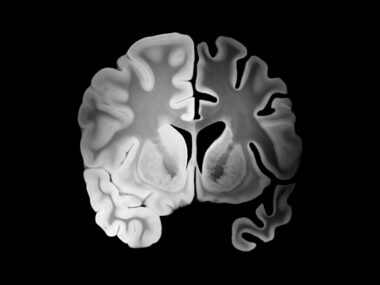
Persistent fatigue in people with multiple sclerosis (MS) is associated with damage in specific areas of the brain, regardless of depression symptoms, a study shows.
The study, “Microstructural fronto-striatal and temporo-insular alterations are associated with fatigue in patients with multiple sclerosis independent of white matter lesion load and depression,” was published in the Multiple Sclerosis Journal.
Fatigue, which affects more than 65% of MS patients, is one of the main causes of reduced quality of life for people with this neurodegenerative disorder. The underlying mechanisms behind MS-related fatigue are still unclear, but disease-associated molecular and cellular processes — including inflammation and brain damage — are among the suggested causes.
Previous studies, which used conventional magnetic resonance imaging (MRI) assessment methods, demonstrated that fatigue is associated with increased T2-enhanced lesion volume — an indication of total brain lesion — and brain atrophy.
Results from diffusion tensor imaging (DTI) also suggested that damage to the frontal lobe — the brain region responsible for voluntary movement, memory, language, attention, and motivation — might play a role in the development of fatigue in people with MS.
DTI is an MRI technique that detects how water travels along the white matter tracts in the brain. It is more sensitive at detecting microstructural changes than conventional MRI methods.
Despite this evidence, other studies have failed to detect significant associations between structural brain damage and fatigue, stressing the need to clarify these potential links. Researchers have said these inconsistent results may be explained by temporal fluctuations of fatigue. That was not considered in the studies, with fatigue assessed only at a single time point.
To learn more, researchers at Harvard Medical School and Washington University School of Medicine in St. Louis evaluated microstructural brain damage in MS patients with different levels of fatigue over time — never, sporadic, or persistent.
They also set out to assess whether fatigue and depression were associated with damage to specific or distinct brain regions. Fatigue and depression — another main cause of impaired quality of life in people with MS — show a considerable overlap of symptoms. However, their relationship is still poorly understood.
The analysis assessed 93 participants from the CLIMB study, an ongoing, prospective, observational study of more than 2,000 MS patients. All participants underwent annual MRI and DTI brain scans, and biennial quality-of-life assessments. Fatigue was measured through the Modified Fatigue Impact Scale (mFIS), and depression through the Center for Epidemiologic Studies–Depression Scale (CES-D).
The participants, of whom 77% were women, had a mean age of 50.6 years, and mean disease duration of 16.9 years.
They were divided according to their fatigue levels over time: 26 had persistent fatigue; 25 had sporadic, reversible fatigue; and 42 had never reported symptoms of fatigue. No significant differences in terms of gender, age, disease duration, and physical disability were found between the three groups.
Results showed that patients with persistent fatigue had significantly higher fatigue and depression scores, as well as overall brain damage, compared with those in the two other groups.
DTI data highlighted that the individuals with persistent or sporadic fatigue had significant microstructural changes in their brains, compared with those without fatigue. The most affected areas were the four cerebral lobes (frontal, temporal, parietal, and occipital), and deep grey matter (mainly made of nerve cells) and white matter (mainly consisting of nerve fibers).
Those abnormalities were more widespread in patients with persistent fatigue than in those with sporadic fatigue.
“Our results suggest that persistent clinically relevant fatigue over years is associated with more extensive brain damage than fluctuating fatigue,” the researchers said.
Further analysis showed that damage of a specific brain network — which connects the cingulate cortex, the postcommissural striatum, the fornix, the corpus callosum, and the thalamus — may be implicated in the development of both fatigue and depression.
Damage to two other brain pathways were specifically associated with fatigue, independent of depression or white matter lesion load. One of these pathways connects the orbitofrontal cortex, subgenual area, and precommisural striatum, and another connects the temporal, insular, and peri-insular regions.
The team noted that understanding the underlying brain mechanisms of fatigue “might pave the way for better predictors of fatigue treatment response and identification of more selective treatment targets for fatigue management.”

Leave a comment
Fill in the required fields to post. Your email address will not be published.