
Cerebrospinal Fluid of MS Patients More Diverse and Filled with Pro-Inflammatory Cells, Study Shows
Written by |

People with multiple sclerosis (MS) have a more diverse set of immune cells in their cerebrospinal fluid (CSF), the fluid that bathes the central nervous system, but no such diversity is seen in their blood, a study reports. Instead, MS causes changes in the activation of immune cells in the blood.
The distinct set of immune cells in MS patients’ cerebrospinal fluid (CSF) shows an enrichment of pro-inflammatory cells that promote disease severity in MS mouse models.
Those findings come from the study “Integrated single cell analysis of blood and cerebrospinal fluid leukocytes in multiple sclerosis,” which was published in the journal Nature Communications.
CSF surrounds the brain and spinal cord (the central nervous system). In healthy individuals, the non-cellular part of CSF is expected to be very similar to that of blood. However, the nature of the cells that compose the CSF is far from completely understood.
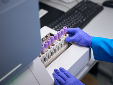
CSF is often sampled — by spinal tap — and analyzed for a diagnosis of MS. That is because it is a site for accumulation of auto-reactive immune cells, including T- and B-cells, that are involved in MS development.
While “evidence supports the involvement of both T- cells and B-cells in MS,” researchers noted, “the relative contribution of each cell type to disease etiology [cause] is unknown.”
Researchers in Germany and California investigated how the cellular composition of CSF changes in MS.
Since the levels of cells in CSF are low, researchers used a cutting-edge genomic technique that identifies unique immune cell types from very small samples.
The team applied single-cell RNA sequencing, a technique that allows the analysis of the transcriptome — the entire set of messenger RNA molecules constituting copies of the DNA sequence used to build proteins — at the level of a unique, single cell.
Patients with idiopathic intracranial hypertension (characterized by increased pressure inside the skull) were recruited as controls in the study, along with naïve-treatment patients with clinically isolated syndrome (CIS) or relapsing-remitting MS (collectively called the MS group). For these groups both blood and CSF samples were collected. (CIS is defined as the first neurological disturbance of MS.)
In total, researchers were able to profile the transcriptomes of 42,969 single blood cells (five controls vs five MS donors), and of 22,357 single cells in the CSF (four controls vs four MS donors).
Results showed that immune myeloid dendritic cells (cells that act as sentinels and can trigger an immune response) and regulatory T-cells (Tregs) were especially enriched in the CSF compared to blood. Tregs act as negative regulators, and like “good police” they shut down the immune response triggered by overreacting T-cells.
By comparing MS samples to controls, researchers saw that MS mainly enhanced the cellular diversity of the CSF, but had no effect on the cellular composition of the blood. Instead the disease changed the transcriptome of immune cells in the blood.
MS altered the immune cells that make the CSF, namely by expanding antibody-producing cells (B-cells), cytotoxic T-cells (especially cytotoxic CD4+ T-cells), and Tregs.
“In blood, MS thus preferentially increased transcriptional diversity, while in CSF it preferentially increased cell type diversity,” researchers wrote.
A more detailed computational analysis, called cell set enrichment analysis (CSEA), showed that T follicular helper cells (also known as Th cell type 1) — known drivers of MS — that support B-cells also were enriched in MS samples.
Then, to validate whether these changes in CSF cellular composition had an impact in MS disease, the team used two MS mouse models, with or without T follicular helper cells.
Results showed that removing T follicular helper cells eased disease severity – mice had fewer inflammatory lesions, and less immune cell infiltrates in the spinal cord. Mice lacking T follicular helper cells also had a lower proportion of B-cells, which drive autoimmunity in MS.
Thus, “such TFH [T follicular helper cells] cells promote CNS [central nervous system] autoimmunity and local B-cell infiltration in two distinct animal models of MS,” researchers wrote.
Based on the results, the team believes that MS is associated with “a pathological interaction between TFH cells and B-cells in the CSF [that] may locally drive CNS autoimmune reactions.”
Overall, the “study provides an essential reference point for … future studies of human CSF, and will likely facilitate understanding of diverse neurological diseases such as Parkinson’s and Alzheimer’s disease,” besides MS, the team suggested.


Leave a comment
Fill in the required fields to post. Your email address will not be published.