
Interaction Between Different Immune Cells May Lead to New MS Therapies, Study Suggests
Written by |

Scientists discovered new interaction between immune cells from the central nervous system (CNS) — consisting of the brain and spinal cord — and immune cells from the blood that may lead to new treatments for multiple sclerosis (MS) and other neurological diseases.
The findings were described in “Microglia response following acute demyelination is heterogeneous and limits infiltrating macrophage dispersion,” a study published in the journal Science Advances.
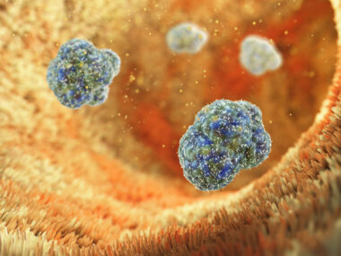
In people with MS, blood immune cells called macrophages produce inflammatory factors that contribute to the deterioration of the myelin sheath — the protective coat surrounding the nerve cells of the CNS. As this process of demyelination goes on, nerve cells become injured.
In the past, it was difficult to tell apart macrophages from microglial cells — immune cells specific to the central nervous system — using conventional techniques. However, in the new study, the researchers used an innovative technique that allowed them to genetically label the two types of immune cells with the goal of telling them apart upon injury to the myelin sheath.
This technique allowed the investigators to find that, soon after the demyelination process started, microglial cells surrounded and encapsulated blood-derived macrophages. This interaction stopped the macrophages from migrating into the demyelinated injury areas.
“We expected the macrophages would be present in the area of injury, but what surprised us was that microglia actually encapsulated those macrophages and surrounded them — almost like police at a riot. It seemed like the microglia were preventing them from dispersing into areas they shouldn’t be,” Jason Plemel, a medical researcher from the University of Alberta, Canada, and the study’s first author, said in a press release.
The team also saw that, over time, microglia cells continued to proliferate while macrophages started to naturally die.
These interactions between the two immune cell types was specific to the CNS, because in injuries that took place in the peripheral nervous system, or PNS — the areas outside the brain and spinal cord — the macrophage population proliferated rather than the microglial population, the researchers found.
Importantly, when the researchers removed microglial cells to see what the role of this interaction was, macrophages moved into uninjured tissue close to the original injury area and caused the loss of nerve cells.
“This suggests that when there is injury, the microglia interfere with the macrophages in our central nervous system and act as a barrier preventing their movement,” Plemel said.
The team demonstrated “that microglia preferentially accrue and monopolize the CNS lesion site, which is in stark contrast to the ongoing accumulation of peripheral macrophages following PNS injury.”
Additionally, the researchers were able to identify at least two different populations of microglial cells. In being able to understand how each type of microglial cell responds to demyelination, they hope to develop new therapies to stop that process and repair damaged nerve cells.
“The indication of at least two different populations of microglia is an exciting confirmation for us,” Plemel said. “We are continuing to study these populations and hopefully, in time, we can learn what makes them unique in terms of function.”
“The more we know, the closer we get to understanding what is going on (or wrong) when there is neurodegeneration or injury, and being able to hypothesize treatment and prevention strategies,” he concluded.

Leave a comment
Fill in the required fields to post. Your email address will not be published.