#ACTRIMS2020 – IL-13 May Be ‘Attractive’ Target for Easing Inflammation in MS
Written by |
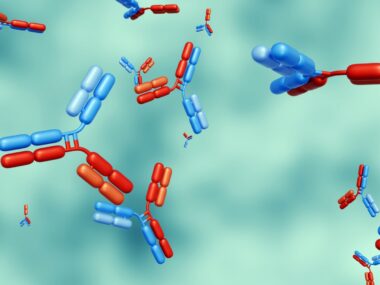
A signaling molecule of the immune system called interleukin 13 (IL-13) may modulate the function of key immune cells involved in multiple sclerosis (MS), and their migration through the barrier that protects the brain and spinal cord.
IL-13 is an “attractive molecule” and a potential avenue for treating MS, researchers said, speaking of its apparent ability to act both on immune cells and their movement into the central nervous system (the CNS; the brain and spinal cord).
These findings were presented by Chloé Hoornaert, PhD, with the CHUM Research Centre in Canada, at the Americas Committee for Treatment and Research in Multiple Sclerosis (ACTRIMS) Forum 2020 held in Florida on Feb. 27–29.
Her presentation was titled “Modulatory Potential Of The TH2 Cytokine Interleukin-13 On The Blood-Brain Barrier And On Immune Cells In Multiple Sclerosis.”
A growing body of evidence has demonstrated a neuroprotective role for IL-13 in multiple animal models of neuroinflammation.
IL-13 belongs to a large group of signaling factors called cytokines — small proteins usually released by immune cells and with a variety of actions, from modulating immune and inflammatory responses to cell adhesion, activation, and differentiation.
Although signaling through IL-13 can be associated with allergic reactions in the airways (e.g., asthma), it can also have important anti-inflammatory properties.
This cytokine can be secreted by various immune cell subtypes, including T helper type 2 cells (Th2 cells; they are typically involved in anti-inflammatory responses), CD4+ T-cells, and natural killer T cells.
In earlier studies using mice with demyelinating lesions — mimicking a hallmark of MS (the loss of myelin, the protective coating of neurons) — Hoornaert and colleagues found that increasing the levels of IL-13 in the brain effectively limited brain lesions and myelin loss.
To determine if these neuroprotective benefits seen in mice might translate to people, researchers studied the cytokine’s effects on immune cells from both MS patients and healthy donors. They also worked on a laboratory model of the human blood-brain barrier — a highly selective membrane that regulates the movement of compounds and cells between the blood and the CNS.
The breakdown of the blood-brain barrier plays a key role in many neurological diseases — including in MS — allowing for activated immune cells circulating in the blood to infiltrate the CNS. There, they trigger the local inflammation that ultimately damages myelin and nerve cells.
Dysregulation of the blood-brain barrier and immune cell migration are, in fact, among the earliest abnormalities seen in the brains of MS patients.
The blood-brain barrier model used in this study employed a layer of human brain endothelial cells and meningeal endothelial cells, both collected from a healthy brain.
Results obtained in vitro (in cells in the lab) showed that the differentiation of T helper 17 (Th17) cells — a group of immune cells involved in inflammatory and autoimmune (self-reacting) responses — boosts production of the IL-13 receptor in a subset of T-cells called CD4+ T-cells.
This means that this T-cell group is now able to detect and respond to IL-13 signals, which is somewhat unusual. A functional IL-13 receptor was long thought to be absent on T-cells.
IL-13 also seems to affect the properties of the blood-brain barrier. Adding IL-13 to the barrier model significantly raised the production of vascular cell adhesion molecule-1 (VCAM-1) — a surface molecule that promotes the binding of certain types of immune cells to blood vessels, a process involved in the migration of these cells to tissues.
This cytokine also enhanced the barrier properties of human brain endothelial cells and meningeal endothelial cells, as evidenced by elevated tight junction components — specialized connections between cells that serve as ‘molecular gates’ to regulate the diffusion of molecules. This enhancement ultimately increased the tightness and integrity of the blood-brain barrier, and reduced its permeability.
According to Hoornaert, “similar beneficial effects” were also observed in human brain endothelial cells derived from MS patients, meaning that cells from patients also responded to IL-13.
These results “demonstrated IL-13R [IL-13 receptor] expression on some key cellular players involved in the [MS] pathological process,” the researchers wrote.
“The capacity of IL-13 to effectively modulate blood-brain barrier function makes this an attractive molecule for further investigation as a potential therapeutic avenue for MS, given its potential to act both at the level of immune cells themselves, as well as on immune cell trafficking across the blood-brain barrier into the inflamed CNS,” the team concluded.