
Abnormal fat in brain immune cells may help drive progressive MS
Study shows enzyme blocking could help reverse activity
Written by |
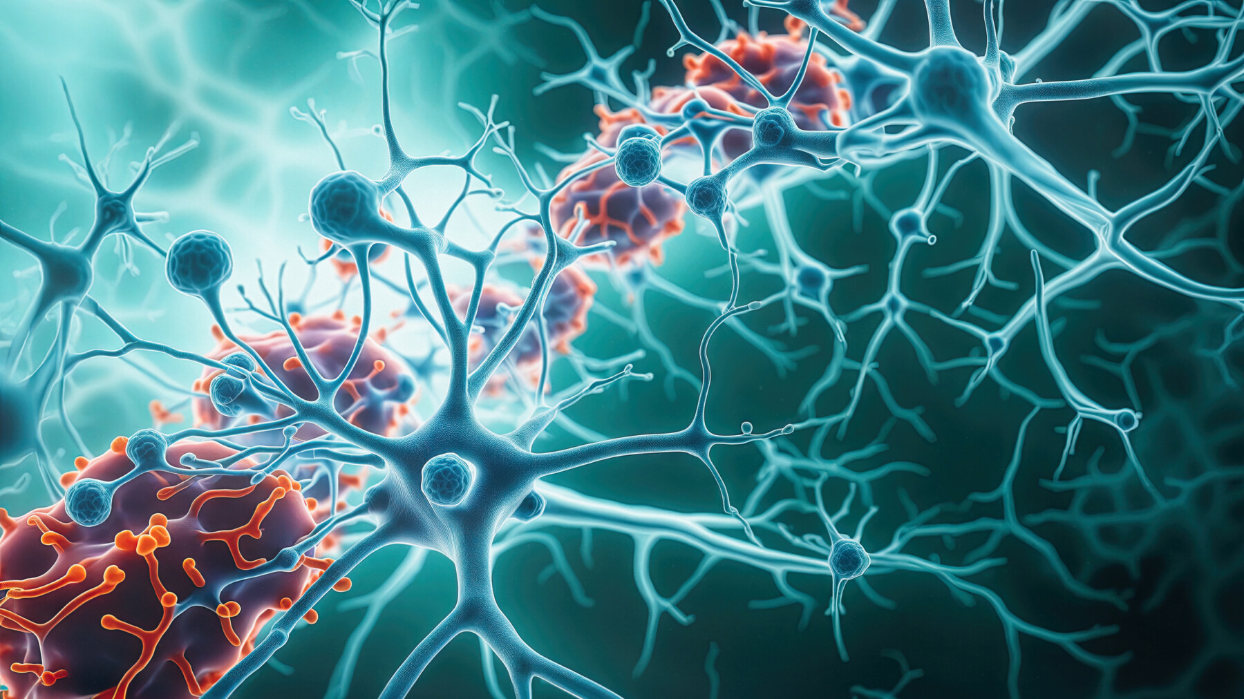
Neurons and microglia work together in the brain. (Image by iStock)
-
Abnormal fat buildup in brain immune cells (foamy microglia) may drive progressive multiple sclerosis.
-
These foamy microglia correlate with faster disability progression in MS patients.
-
Blocking the MAGL enzyme could reverse this activity, offering a potential new treatment target.
Abnormal fat buildup inside the brain’s immune cells may help to drive neurological damage and disability progression in people with progressive forms of multiple sclerosis (MS), a study found.
The findings also point to a possible therapeutic target. Researchers showed that blocking an enzyme called MAGL may help reverse the disease-driving activity of these abnormal immune cells. A treatment targeting this enzyme is in clinical development as a potential MS treatment.
The study, “Foamy microglia link oxylipins to disease progression in multiple sclerosis,” was published in Nature Neuroscience. Microglia are immune cells in the brain that normally play key roles in protecting the brain from infections and other threats. These cells are also important for clearing waste and helping repair damaged tissue. In MS, however, microglia can become chronically activated and contribute to inflammation.
MS is a chronic disorder marked by damage to the myelin sheath, a fatty covering around nerve fibers, the brain, and the spinal cord, which shows up on MRI scans as lesions. Over time, some of these lesions resolve, while others continue to expand, indicating ongoing tissue damage.
While scientists know that slowly expanding brain lesions are linked to MS progression, the biological mechanisms driving this ongoing damage have remained unclear.

‘Overloaded’ cells can no longer do their job
The scientists conducted detailed analyses of brain tissue from deceased donors with secondary progressive MS (SPMS) to examine whether microglia may contribute to the gradual expansion of lesions in MS.
Results showed that some MS lesions — particularly those that showed signs of active inflammation — were filled with microglia that contained many abnormal fatty deposits. These cells are known as foamy microglia, because under a microscope the fat deposits resemble bubbles or foam.
The researchers found that patients with a greater proportion of lesions containing foamy microglia had generally experienced faster disability progression during life.
“We found that patients with large numbers of these foamy microglia had a more severe disease course more frequently,” Daan van der Vliet, study lead author and a researcher at the Netherlands Institute for Neuroscience, said in an institute news story. “These cells are probably trying to do something good: clearing up damage. But they become overloaded, so to speak. As a result, they can no longer effectively contribute to repair.”
The scientists stressed that these analyses were limited to tissue from deceased donors, making it impossible to draw conclusions about how these cells behave in living patients. Nonetheless, they said, the data suggest that foamy microglia may contribute to disability progression.
“Across 250 MS donors, we found that the proportion of lesions with foamy microglia correlated with faster progression to [worse disability] scores, while lesions with nonfoamy microglia were not associated with fast disease progression,” the researchers wrote. “This finding may suggest that the foamy phenotype is not simply a byproduct of [nervous system damage] but a defining feature of lesion persistence and expansion.”
Comprehensive analyses of the biochemical activity of foamy microglia in MS lesions then found that these cells had increased MAGL activity. This enzyme breaks down fatty molecules into signaling molecules called oxylipins, which help regulate inflammation and cell-to-cell communication.
Spurred by this finding, the team tested the effects of blocking MAGL in a mouse model of MS-like brain damage. This led to signs of neurological repair that are normally not seen in this model.
“These findings suggest that targeting MAGL may offer a disease-modifying approach for MS progression by promoting lesion resolution and repair,” the researchers wrote, adding that experimental therapies targeting MAGL are being tested in clinical trials.
The study also identified potential biomarkers of foamy microglia. Levels of oxylipins in cerebrospinal fluid — the liquid surrounding the brain and spinal cord — closely matched the proportion of foamy microglia in patients’ lesions.
“That opens the possibility of developing biomarkers in the future that could help doctors identify earlier which patients are at risk of rapid decline — and which treatment would suit them best,” van der Vliet said.


Mark Collins
Hopefully this research will go at lighting speed we need help now