
Study uses lab-grown ‘mini brains’ to explore myelin repair in MS
It's one of three projects to receive funding from MS Australia
Written by |

Three initiatives have been funded through MS Australia’s 2026 Mid-Year MS Research Grants Round. (Image from iStock)
- Australian researchers are using lab-grown "mini brains" to investigate myelin repair in multiple sclerosis (MS).
- Other studies explore the role of the Epstein-Barr virus in MS and spinal cord imaging.
- These projects aim to advance understanding and develop new treatments for MS.
Australian researchers are using lab-grown “mini brains” to investigate new ways to repair myelin, the protective coating surrounding nerve cells that is lost in multiple sclerosis (MS).
The 3D lab models, grown from human stem cells, will allow researchers to study how myelin forms naturally in the human brain.
The project, funded by MS Australia, is led by Samantha Barton, PhD, at The Florey Institute of Neuroscience and Mental Health.
“We’ve developed laboratory-grown brain models that allow us to study how human myelin forms in ways that aren’t possible using conventional laboratory techniques or animal models,” Barton said in an MS Australia press release.
Current treatments do not repair myelin damage
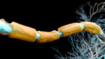
In MS, inflammatory attacks damage myelin, a substance mainly made of fatty molecules and specialized proteins that helps nerve fibers in the brain, spinal cord and optic nerve efficiently send the electrical signals needed for proper neurological function.
The body can repair or replace myelin through a process called remyelination. However, this repair becomes less effective as age advances. Also, while existing disease-modifying treatments for MS can lower the risk of relapses and slow disease progression, ways to promote remyelination is an active area of research.
“Current treatments target the immune system, but they can’t repair the damage that has already occurred,” Barton said.
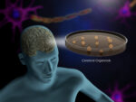
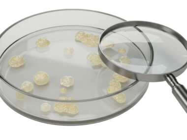
Barton’s team will use cerebral organoids, or “mini-brains,” which contain cells organized in 3D resembling how they would appear in the human nervous system. This will give the researchers a better look at the genes and molecular pathways involved in forming myelin.
“By understanding the genes and biological pathways involved in myelin formation, we hope to discover new ways to promote myelin repair,” Barton said. “As a researcher, it’s incredibly rewarding to ask questions that have never been answered before.”
The project addresses a research area that people living with MS have identified as particularly important, according to Fiona McKay, PhD, MS Australia’s deputy head of research.
“When we asked people affected by MS which research areas mattered most to them, repair and regeneration emerged as the top priority,” McKay said. “Research like Dr Barton’s is improving our understanding of how myelin forms and how it may one day be repaired. Supporting discovery research like this builds the knowledge needed to develop the next generation of treatments for people living with MS.”
Rohan Greenland, CEO of MS Australia, said continued investment is essential to advancing MS care.
“MS Australia is committed to supporting Australia’s leading researchers as they tackle some of the biggest unanswered questions in MS,” Greenland said.
Other projects target Epstein-Barr virus, spinal cord damage
Barton’s project is one of three initiatives funded through MS Australia’s 2026 Mid-Year MS Research Grants Round. The funding round includes two Incubator Grants, intended to help researchers generate preliminary evidence for larger research programs, and a postgraduate scholarship for the next generation of MS scientists.
A second project, led by Carla Proietti, PhD, at the University of Queensland, will investigate how inherited genetic factors play a role in the immune response to Epstein-Barr virus (EBV).
EBV, a member of the herpes virus family, has been increasingly identified as a major environmental risk factor for MS. Notably, while almost everyone is infected with EBV, virtually all people with MS have had a past EBV infection. This suggests that this virus interacts with other risk factors to initiate the MS disease process. Proietti’s team will combine genetic analyses with detailed assessments of antibodies targeting EBV and human proteins to better understand the link between this virus and MS.
“We know Epstein-Barr virus is one of the strongest risk factors for MS, but we still don’t understand why only some people go on to develop the disease,” Proietti said.
The researchers hope their findings will support earlier diagnosis, more personalized care, and the development of better treatments.
By improving how we image the spinal cord, we hope to better understand how spinal cord damage contributes to disability over time.
The third project, led by Tal Koren, MD, at the University of Sydney, will use advanced MRI scans and artificial intelligence to improve the detection and tracking of spinal cord damage in people with MS.
More accurate imaging could help clinicians monitor disease progression and determine whether a treatment is working or should be changed.
“By improving how we image the spinal cord, we hope to better understand how spinal cord damage contributes to disability over time,” Koren said. “This could help clinicians monitor disease more accurately, identify people at greater risk of progression, and uncover new biological pathways that may become future treatment targets.”


John Cruz
My God I'm so excited I can't believe Dr. Onokun could cure multiple sclerosis i had for 3year using healing herbs! This works like magic to me, I'm forever grateful He can also cure any kind of disease or virus. contact him via: onokunherbs.com