
Scientists link early relapsing MS to subtle changes in brain white matter
Specialized technique reveals damage despite 'normal' MRI findings
Written by |
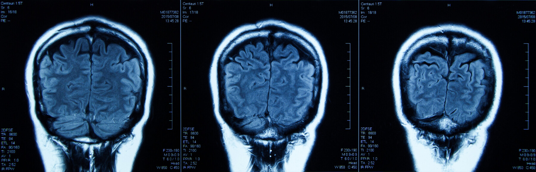
Researchers used MRI scans and a specialized technique to investigate white matter in the brain. (Image from iStock)
- Scientists found subtle changes in normal-appearing brain white matter on MRI scans of people with early relapsing-remitting multiple sclerosis.
- Using a specialized noninvasive imaging technique, the team identified metabolic abnormalities in the brains of RRMS patients.
- The findings are preliminary, however, and require validation through larger, long-term studies before becoming clinical disease biomarkers.
A trio of European scientists has linked subtle changes in the brain’s white matter — tissue rich in nerve cell projections — to early relapsing-remitting multiple sclerosis (RRMS), a form of the neurodegenerative disease marked by flare-ups interspersed with periods of remission.
Using a specialized noninvasive imaging technique, the team identified metabolic abnormalities in the white matter that had appeared normal on standard MRI scans done among people with RRMS in its early stages, according to a new study.
These changes in white matter varied by brain region and generally reflected mixed patterns of tissue injury, the researchers found. Certain changes correlated with aspects of cognitive function in the RRMS patients, though the researchers noted that most of these relationships were relatively weak and likely influenced by other variables. Meanwhile, the relationships to a mixed measure of physical and cognitive function were stronger, the team noted.
Taken together, the findings support metabolic injury in normal-appearing white matter as “a genuine feature of early disease,” while suggesting that its relationship with cognition is “modest and confounded,” the researchers wrote. The trio emphasized that these results — showing “associations [that] are detectable but weak” — are preliminary and require confirmation in larger, long-term studies.
The study, titled “Regional normal-appearing white matter metabolic profiles and cognition in early relapsing–remitting multiple sclerosis: a multi-voxel proton magnetic resonance spectroscopy study,” was published in the journal Acta Neurologica Belgica.

A chronic condition, multiple sclerosis (MS) is caused by an immune system attack that damages myelin, the protective coating around nerve fibers. In the brain and spinal cord, white matter is the tissue composed mainly of myelinated nerve fiber projections, which carry electrical signals throughout the brain.
In MS, cognitive problems are common but variable. Moreover, the amount of visible brain damage on conventional MRI scans does not always correlate well with the severity of cognitive dysfunction. One possible reason for this, according to the researchers, is that standard MRI can miss more subtle microscopic signs of injury.
Differences seen in brain white matter of patients vs. healthy people
Brain white matter that appears normal on a standard MRI can be examined in more detail using proton magnetic resonance spectroscopy, or 1H-MRS. This noninvasive imaging technique measures certain chemicals in brain tissue to better understand its metabolic activity.
Here, the researchers sought to determine whether 1H-MRS could detect subtle white matter abnormalities in people with early RRMS who had normal-appearing white matter on MRI scans. The trio also evaluated whether these changes were associated with cognitive function.
Their analysis involved 51 adults with early RRMS and relatively mild disability, as well as 44 healthy people, who served as controls. In both groups, 81% of participants were women, and the mean age was approximately 40. Each MS patient was being treated with interferon-beta.
The scientists used 1H-MRS to examine six white matter regions throughout the brain that had looked normal on MRI. These included the frontal and parietal areas, and deep regions on both the brain’s left and right sides.
The team found that, in four of the six areas, an indicator of nerve fiber health — known as the N-acetylaspartate-to-creatine ratio or NAA/Cr — differed between MS patients and healthy controls.
People with RRMS had lower NAA/Cr in the left frontal, deep, and parietal white matter, consistent with reduced nerve cell health. Unexpectedly, the MS patients had a higher NAA/Cr in the right deep white matter — though the researchers believe this reflected technical factors rather than a true biological difference.
The choline-to-creatine ratio was elevated in four of the six regions among people with MS, a pattern consistent with increased breakdown of cell membranes and myelin loss. Changes in the myo-inositol-to-creatine ratio, which reflects the activity of nerve support cells called glia, varied by location, further demonstrating that metabolic abnormalities were not uniform throughout the brain, the team noted.
In healthy brain tissue, NAA/Cr shows a gradient, with the lowest levels at the front of the brain and increasing toward the back. This gradient was generally observed in both healthy individuals and MS patients, but its configuration was significantly altered in the MS group, the researchers noted. That finding supports the idea that RRMS reshapes regional metabolic patterns in the brain, per the scientists.
The researchers then examined whether metabolic white matter changes were associated with cognition in the individuals with MS.

Findings, though, are weak and require further study
Initial analyses found that a higher NAA/Cr in the left frontal region was associated with faster information processing, while higher levels in the left parietal region were associated with better verbal learning. However, in final analyses that adjusted for other variables, NAA/Cr was not independently associated with these cognitive measures.
In contrast, NAA/Cr in several regions — particularly the frontal white matter — was significantly and independently associated with scores on the Multiple Sclerosis Functional Composite, a broader measure that combines walking, arm and hand function, and cognitive processing.
No metabolic measures were associated with fatigue or mood, the team noted.
Overall, “early RRMS shows regionally heterogeneous [dissimilar] … metabolic change,” the researchers wrote. The team noted that the links between NAA/Cr and purely cognitive measures were weak, but were stronger in mixed measures of physical and cognitive function.
Among the study limitations were its relatively small size and the fact that patients were only assessed at a single point in time. Using that single time point prevents determining whether these metabolic abnormalities occur before or after cognitive changes.
Larger studies following patients over time will be needed before these measures can be considered potential disease biomarkers, the team noted. Still, the researchers say these findings are “hypothesis-generating.”



Michele Chadwick
This is consistent with my MS. I was dx at 28, symptoms since teens. I don’t have many physical symptoms. But have always had cognitive symptoms even as a teen. I am about to be 57 and on disability now and is a lot easier without work pressure. But I do have some mental health issues, depression and anxiety. I thought in the beginning I would lose the ability to walk , but never knew that these cognitive problems are MS too is scarier. I am still intelligent, but processing is very slow and word recall is terrible. Add that to age and progression and mild physical symptoms are becoming known. This is a terrible disease. I have been on a disease modifier from time of dx.