
Study of Myelin-producing Cells a Step Forward for MS, Other Neurological Disorders
Written by |
A study found that the cells responsible for the production of myelin selectively introduce a myelin-insulating layer in a particular set of neuronal axons in the brain’s white matter.
This represents a step forward in the basic mechanisms that may underlie neurological disorders, including multiple sclerosis (MS). Also, a newly developed method to visualize these cells will help scientists investigate demyelinating diseases.
The study, “Rabies virus-mediated oligodendrocyte labeling reveals a single oligodendrocyte myelinates axons from distinct brain regions,” was recently published in the journal Glia.
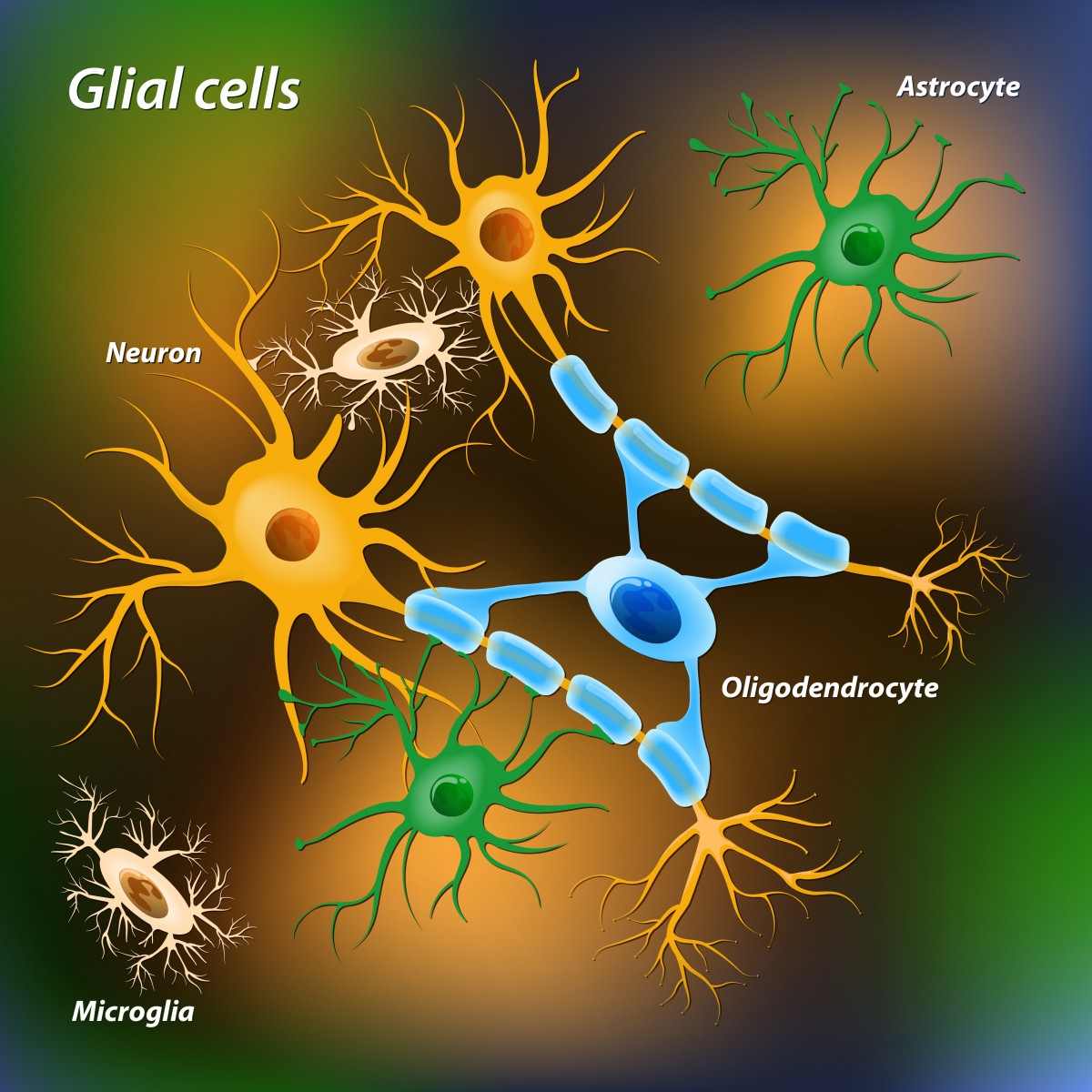
Glial cells are a type of cell found in the central nervous system. Although they are not neurons, they have crucial functions: maintaining homeostasis, forming myelin, and providing support and protection for neurons.
In the brain there are three kinds of glial cells: oligodendrocytes, astrocytes, and microglial. The major function of oligodendrocytes is to form myelin, the insulator layer protecting neuronal axons and increasing the velocity of neuronal impulses.
An axon is a long, slender projection of a neuron whose job is to transmit information (electrical impulses) to different neurons, muscles, and glands. Neuronal axons extend from multiple brain regions and pass through the brain’s white matter.
Scientists have questioned whether oligodendrocytes enclose a particular set of axons or do so randomly in our brains.
In this study, a Japanese research team at the National Institute for Physiological Sciences (NIPS) in Okazaki, Aichi, Japan, developed a new strategy to visualize individual oligodendrocytes and axons derived from particular brain regions in the white matter of mice.
Using this method, researchers simultaneously labeled neuronal axons derived from distinct brain regions (motor cortex or sensory cortex) and oligodendrocytes in the white matter. They found that oligodendrocyte action for myelination is not solely random, but some oligodendrocytes selectively myelinated axons from a particular brain region.
These results represent an important step in understanding the interaction between oligodendrocytes and neuronal axons. This is especially relevant for severe neurological disorders, such as MS, where oligodendrocyte dysfunction is known to contribute to disease severity.
The new method will assist scientists in investigating demyelinating diseases.
“Now, we plan to analyze oligodendrocyte morphology and myelination in demyelinating mouse models,” Dr. Takeshi Shimizu, the study’s lead author, said in a press release. “Furthermore, axon selective myelination for a specific neuronal subtype found in this study encourages us to investigate physiological relevance of multiple myelination to higher brain function.”


Leave a comment
Fill in the required fields to post. Your email address will not be published.