
Thin But Persistent Regrowth of Myelin Layers Sign of Health in CNS, Study Says
Written by |

The generation of a thin myelin sheath during remyelination — one that continues to protect nerve cells over time — is indicative of the long-term health and activity of the central nervous system (CNS) in demyelinating diseases such as multiple sclerosis (MS), a new study shows.
These findings, which aim to settle a scientific debate about CNS healing, are reported in the study “Thin myelin sheaths as the hallmark of remyelination persist over time and preserve axon function,” published in the journal PNAS.
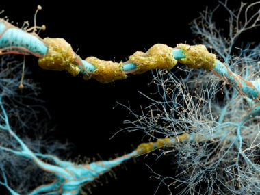
The myelin sheath is an insulating layer of lipids (fats) and proteins that wrap around nerves. It works to speed the transmission of signals from the nerves to muscles, in order for a person to carry out activities such as walking, talking, and breathing.
Remyelination, which refers to a process through which the body rebuilds the myelin sheath when it has been stripped away due to disease, is the most powerful form of repair for the brain and the spinal cord in patients with a demyelinating disease. In the recovery phase of a disease, the body will completely remyelinate areas that have been stripped of myelin.
MS is a demyelinating disease, and in these patients remyelination can be quite extensive. The ability of the body to remyelinate nerve cells can also decline as a person ages.
Remyelination is characterized by the presence of thin myelin sheaths. Diameter wise, these new myelin sheaths are thinner than the original myelin layers. While some studies suggest that the myelin layer eventually returns to the original diameter, others suggest that it stay thin.
Researchers at the University of Wisconsin-Madison believe that remyelinated sheaths remain thin, and previously described observing predominantly thin myelin sheaths in a canine with a demyelinating disorder. But this view remains controversial.
The team, for this reason, conducted a long-term study of two animal models of demyelination.
The first model, studied for more than a decade, was a dog with a genetic developmental delay in myelination. The dogs formed thin myelin sheaths in ways similar to changes seen in remyelinated nerves.
Researchers showed that in this animal model the myelin sheaths remained thin and stable on many axons (nerve cells projections) during the dog’s lifetime, and that these thinner-than-usual myelin sheaths had no detrimental effects on the axons’ ability to transmit nerve impulses or signals.
Next, they studied cats that were undergoing remyelination due to a demyelinating disease. In this animal model, there was significant demyelination of the spinal cord and the optic (eye) nerves. The researchers followed these cats for two years, and demonstrated that the thin myelin sheaths persisted over this period.
“We found that nearly every optic nerve fiber was remyelinated with a thin myelin sheath, which is important for understanding human disease because in multiple sclerosis, the optic nerve is often the first to be demyelinated,” Ian Duncan, the study’s first author, said in a news release.
Overall, researchers demonstrated the importance of remyelination via the production of thin myelin sheaths in the long-term health and function of the CNS. More significantly, they confirmed that the best way of evaluating the health of remyelination and its central nervous system benefits is the persistence of thin myelin sheaths.

GARY SHAMBLEN
Great. that's what all us MSers need yo know for our dog or cat. All joking aside, what can we MS patients do toward achieving remyelination?
Tim Pruitt
I’m interested in knowing how to, as well...
Holmes
I am interested too. How are they achieving this???
huri tursan
torturing mice, cats, dogs by dehumanizing them by calling them 'models'. Not in my name as I totally disapprove of vivisection as this type of research is nothing but vivisection and instead of boasting of results it should not be allowed.
Suzanne Hughes
I don't like the idea of using animals for testing, but it gives scientists a basis to see what will work and what won't work. Did you want to offer yourself to scientists to test on? I think not, haha!
Selina
Yess totally agree. Leave the animals alone!
Billy
I totally approve scientific research that can help my debilitating disease... How dare You? If you could walk a day in my shoes
Sherry Leighton
Having ms is no joke. I have a support group on Facebook called MS Stories. Please feel free to join and share your story. Love n light to you all. ??
Sam Bennett
Your disapproval is noted and summarily dismissed. This is excellent news that offers some real hope for those of us saddled with MS. Now to find a good (and affordable) treatment to promote relational.
Tim Pruitt
How does one achieve this for themselves?
Larry s
When will this be done to people?
susan jo miller
How close are we for MS patients
Josie
Vitamin D actually does help to remyelinate!! Check out the Coimbra Protocol!! Also check out Progesterone to assist with remyelination. The research on Progesterone is old, but it's out there!!
Erik
Biotin, lions mane mushroom and clemastine Fumarate are theorized to stimulate remyelination.
Gail Williams
This sounded so good and hopeful until I read the part about age haviing a big factor for this. Figures everything else is not helping. Sorry for being a downer.
haslie kemp
Not for me either since I am 78 and have had MS for 35 years or more. But one thing my Doctor said is that the older I get my immune system in not as active and not attacking me nervous system and she call it "leveling off"
Albert Matarazzo
I am suffering from MS. Everything I read about MS is in the research stage, or it worked on a rat. I have been going to the same doctor for six years now every three months. Every time I read about stem cell treatment or a new Trial or remyelination I say how about me and I am always met with a negative response. I am on the internet every day looking for any new developments related to MS. I am tiered of looking for false hope. I don't think there will be any major developments in my lifetime so I'm going to just manage my pain as best I can, and pray for a cure for future generations.
MargaretR
Another drug for the same thing (RRMS) is more profitable for drug companies than testing a new drug for remylenation that would help all of us.
Mark S.
That’s the plan. I don’t like being negative but the truth is no cure. Maybe someday a therapy for remylenation. Not sure if that will help with function by the time we get there.
Carol Trautt
I'm 68 and been on Copaxone almost 4 yrs since my diagnosis. (I had MS before that, but blamed my symptoms on other things.) It has kept me stable and may have even helped a little. I read about taurine and clemastine fumarate studies. I'm adding them into my regimen. While I do this I'm going off my low dose statin as cholesterol is needed for a building block for myelin sheath. Besides all the statins I've been on give me leg cramps. I also take Vitamin D and several other supplements.
Ken
I just started using Taurine, but did not know about low dose statins, which I take thanks for the info.
Katya
Me too. Go ahead ! Will see the results after 3-6 months.
Carol Welsh
Sounds very much like my situation. Diagnosed nearly six years ago at 61. On Copaxone and baclofin . No new leisons since diagnosis but pain increasing and mobility more and more a problem. Now trying medical marijuana with no real effect. Want to try taurine and cllemastine, how and how much?
Patricia
What other supplements do you take?
Anthony Crandall
Until nano procedures can assist in any treatment. These all sound like wonderful things in the future. But most people that suffer and I mean suffer from prolonged issues of MS don't really have issue wit DE or remylenation problems. It is scar tissue that block signal use. Remove it and then a way for the body to repair the nerve is what more people that see these reports want. They have hope things will work to improve their lives even more than help those in the future. Show promise of today and the line will be long for test subjects I am sure.
Andrea
On copaxone the burning hiving injection diagnosed 1year Ago at 54 Is it working MRI after MRI thickening of lesions. WTF.