
Enrollment in MS imaging study reaches halfway mark
Mass General, Quantum test PET imaging tracer to measure myelin damage
Written by |
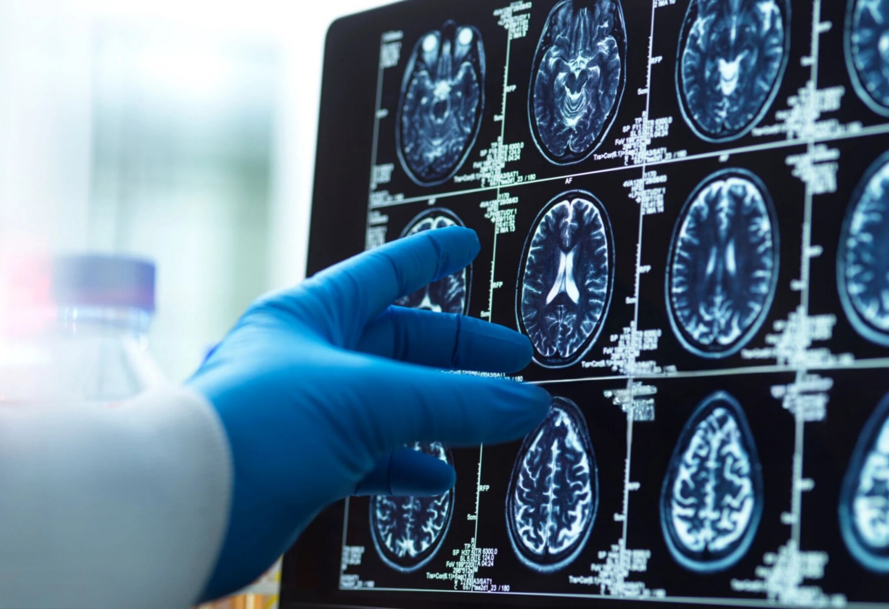
A brain scan generates images for doctors and researchers to analyze. (Photo by iStock)
- A study is testing a PET imaging tracer to detect myelin damage in multiple sclerosis.
- This tracer could provide a direct, quantitative measure of myelin loss and repair in MS patients.
- It aims to improve monitoring of therapies that target myelin protection and restoration.
Enrollment is half complete in an ongoing clinical study at Massachusetts General Hospital (MGH) testing whether a PET imaging tracer can reliably measure myelin damage in people with multiple sclerosis (MS).
The Phase 1 study (NCT04699747) is recruiting about 60 people — healthy adults and MS patients — who will receive the [18F]3F4AP tracer before undergoing PET imaging. The goal is to determine the tracer’s safety and pharmacological properties, and whether it can effectively detect areas of myelin loss (demyelination) in the brain.
The trial is being conducted in collaboration with Quantum Biopharma, which is developing an oral therapy called Lucid-MS that aims to slow myelin damage and potentially reverse some of the nerve damage seen in MS patients. If validated, the new tracer could help more precisely monitor, in real time, how Lucid-MS affects myelin loss and repair.
Preliminary data from the first group of trial participants have shown that the tracer can detect acute MS lesions, and may also identify lesions in gray matter (brain regions consisting mainly of nerve cell bodies).
“PET imaging with [18F]3F4AP has the potential to fundamentally change how we assess demyelination—providing a direct window into [nerve fiber] health and enabling us to more clearly demonstrate the impact of therapies like Lucid-MS that aim to protect and restore the myelin sheath in MS,” Andrzej Chruscinski, MD, PhD, Quantum’s vice president of scientific and clinical affairs, said in a company press release.

Pinpointing demyelinated nerve fibers
In MS, the immune system mistakenly attacks myelin, a protective coating around nerve fibers that helps them transmit electrical signals efficiently, in the brain and spinal cord. Damage to myelin disrupts normal nerve signaling and ultimately drives MS symptoms.
While imaging techniques such as MRI scans are commonly used in MS to detect neuronal damage, they generally cannot precisely measure myelin changes to assess how patients are responding to newer drugs such as Lucid-MS that aim to prevent myelin damage.
[18F]3F4AP is a radioactive form of dalfampridine, the active ingredient in Ampyra (dalfampridine), an approved MS therapy designed to improve walking ability. Dalfampridine can bind to potassium channels on the surface of nerve fibers that become exposed when myelin is damaged, and researchers believe the radioactive tracer may highlight demyelinated nerve fibers during PET scans.
The tracer demonstrated high sensitivity for detecting demyelinated lesions and a favorable pharmacological profile in previous preclinical and clinical studies, making it a promising biomarker for assessing myelin loss and repair.
In the ongoing study, participants undergo PET scans after receiving a rapid infusion of the tracer lasting about one minute. The scanner will show which brain regions are taking up the tracer, with regions that lost more myelin expected to take up a larger amount and appear brighter on the scans. Researchers will compare PET images with MRI scans to determine how accurately the tracer identifies demyelinated lesions.
“From a scientific perspective, the ability to directly quantify demyelinated lesions with intact [nerve fibers] in vivo represents an important unmet need in multiple sclerosis research,” said the tracer’s developer, Pedro Brugarolas, PhD, an investigator at MGH and an assistant professor at Harvard Medical School. “If further validated, this imaging approach could provide a more direct and quantitative measure of myelin loss and repair, which may help improve the evaluation of disease mechanisms and therapeutic response in MS.”
Quantum is planning a Phase 2 trial to assess the efficacy, safety, and tolerability of Lucid-MS in people with MS. The company has submitted an application to the U.S. Food and Drug Administration seeking clearance to start the trial.


Teresa Crisafulli
Interesting study