
AI partnership aims to sharpen brain scans’ detection of MS
Neurophet venture to develop algorithms to spot lesions
Written by |

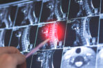
Scientists use AI to help study the brain. (Image by iStock)
- A partnership will develop AI software to improve MS diagnosis.
- It will detect specific brain lesions (CVS, PRLs) on MRI scans, crucial for diagnosis.
- The partnership aims to enhance the clinical reliability of AI algorithms for MS.
A new partnership aims to advance software that uses artificial intelligence (AI) to examine brain MRI scans for signs characteristic of multiple sclerosis (MS).
Neurophet said it will work with the Vall d’Hebron Institut de Recerca (VHIR), the research institute of Vall d’Hebron University Hospital in Spain, to develop algorithms that can detect lesions with the central vein sign (CVS) and paramagnetic rim lesions (PRLs), two MRI features that can be used to support an MS diagnosis under the most recent diagnostic criteria.
VHIR will provide high-quality MRI datasets from people with MS, along with lesion annotations provided by specialists. Neurophet will use the data to develop and validate new AI algorithms for its Aqua MS software, which the U.S. Food and Drug Administration (FDA) has cleared to identify other MS-related changes in brain scans.
“This collaboration with VHIR enables us to directly develop and validate Neurophet’s AI-based multiple sclerosis analysis technology through a leading institution in Europe,” Donghyeon Kim, PhD, co-CEO of Neurophet, said in a company press release. “Going forward, we plan to further strengthen the clinical reliability of our AI algorithms based on large-scale datasets.”
MRI scans are widely used to diagnose MS and monitor disease activity. They can reveal lesions caused by damage to myelin, the protective coating around nerve fibers, as well as brain shrinkage, which can occur as the disease progresses due to neurodegeneration.
Zeroing in on lesions
Neurophet Aqua MS is a module within the company’s Neurophet Aqua brain imaging analysis platform that analyzes MRI scans for features associated with MS. The FDA cleared it to analyze brain atrophy and to identify brain lesions caused by MS, allowing doctors to track changes in lesion number and size over time.
To establish an MS diagnosis, doctors will look for evidence that disease activity has affected the brain and spinal cord in characteristic ways. The CVS, which refers to a small vein visible in the center of an MS lesion, and PRLs — lesions surrounded by a rim of iron-containing immune cells that indicate chronic, ongoing inflammation — are two patterns of lesions common in MS but rare in other conditions.
According to the most recent updates to the McDonald criteria — a set of internationally recognized guidelines for diagnosing MS — these lesions can support an MS diagnosis when used alongside other diagnostic features. The collaboration will focus on training and validating AI models to automatically detect CVS and PRLs in MRI scans.


Leave a comment
Fill in the required fields to post. Your email address will not be published.