
Could Myeliviz, a New Imaging Agent of Myelin, Be a Game-changer?
Written by |

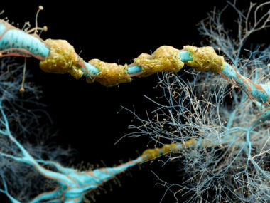
Myelin is the protective sheath that covers nerve fibers and is damaged in those with multiple sclerosis. Quantifying the degenerative process of myelin would lend perspective to how much and where a patient is progressing.
Currently, MRIs are used for diagnostic purposes, but the nuances of progression remain difficult to trace. What if we could track demyelination? This could help narrow the gap between diagnosis and relapse-causing disability. What if we could see, and ultimately repair, that process? What if we could eliminate the chasm?
According to a recent report in Multiple Sclerosis News Today, researchers at Case Western Reserve University have created a new imaging agent for myelin called Myeliviz. The U.S. Food and Drug Administration has since given the green light for Myeliviz to be evaluated in a clinical trial with healthy volunteers. In addition to aiding the diagnosis of MS, Myeliviz could also help to diagnose and track other myelin-associated diseases, as well as their progression.
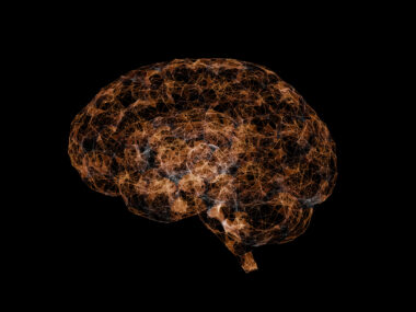
“Myelin has never been directly imaged before,” Yanming Wang, PhD, Myeliviz’s co-inventor and a professor of radiology at Case Western Reserve School of Medicine, said in a press release reported by Multiple Sclerosis News Today‘s Marta Figueiredo. “Our technique is the first to do so and we are hopeful this will provide an earlier and more accurate diagnosis of MS.”
Injected intravenously, Myeliviz binds to myelin and can be seen via positron emission tomography, or PET scans. Myelin is subsequently “lit up,” with areas of myelin loss characterized by low or absent light, according to the report. Preclinical studies in rodents have shown promising results. Many of us with MS could embrace something like this that shows promise.
MRIs have long been my only guidepost while living with secondary progressive multiple sclerosis. They highlight which lesions are active, yet they are often prescribed after a disability has occurred. My last MRI showed new, active lesions in my cervical spine only after I began experiencing exacerbated foot drop and dysphasia. Myeliviz has the potential to offer insight in real-time. Let’s take it one step further. Imagine having the capability to prevent disability with this intervention?
Robert Fox, vice chair for research at the Cleveland Clinic’s Neurological Institute, said, “the goal is to enable clinicians to more unambiguously diagnose MS and monitor disease progression and repair processes,” the report noted. Myeliviz is ready for a human trial at the Cleveland Clinic Mellen Center for Multiple Sclerosis. That data will then be compared with conventional MRIs. Chunying Wu, Myleliviz’s co-inventor and instructor of radiology at Case Western, suggests Myliviz could be the missing link in finding a cure for MS.
Let’s hope. Myeliviz could be the first imaging agent to offer a real-time observation of demyelination. That would be a game-changer — in a game I plan to win.
***
Note: Multiple Sclerosis News Today is strictly a news and information website about the disease. It does not provide medical advice, diagnosis, or treatment. This content is not intended to be a substitute for professional medical advice, diagnosis, or treatment. Always seek the advice of your physician or other qualified health provider with any questions you may have regarding a medical condition. Never disregard professional medical advice or delay in seeking it because of something you have read on this website. The opinions expressed in this column are not those of Multiple Sclerosis News Today or its parent company, Bionews Services, and are intended to spark discussion about issues pertaining to multiple sclerosis.

Leave a comment
Fill in the required fields to post. Your email address will not be published.