
Blood NfL Potential Marker of MS Therapies’ Effectiveness, Study Suggests
Written by |
Starting treatment with a disease-modifying therapy (DMT) reduces blood levels of neurofilament light chain (NfL) — a potential biomarker of disease progression and activity — to varying degrees depending on the therapy used, according to a large real-world study of patients with relapsing-remitting multiple sclerosis (RRMS).
The findings support the potential of plasma NfL as a treatment response marker, useful for monitoring the effectiveness of MS therapies and see how well they are working for patients, researchers said.
The study “Blood neurofilament light levels segregate treatment effects in multiple sclerosis,” was published in the journal Neurology.
Neurofilaments are nerve cell-specific components that can be released upon nerve cell damage or death (neurodegeneration). Neurofilament light chains (NfL) can be measured in different body compartments, and have emerged as promising biomarkers of neurological diseases, including MS.
NfL levels above a certain threshold in the cerebrospinal fluid (the fluid that bathes the brain and spinal cord) or in blood have been proposed as potential biomarkers of MS progression, disease activity, and response to treatment.
The potential value of NfL as a marker for MS “is especially high,” researchers wrote, since it may detect changes over shorter periods of time that are beyond reach through standard magnetic resonance imaging (MRI).
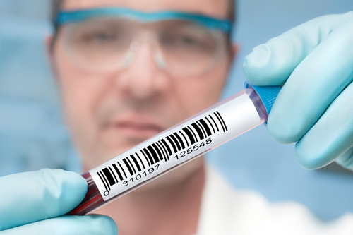
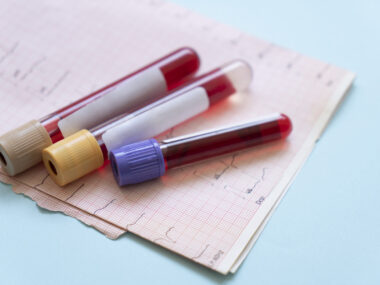
Most studies testing NfL as a marker of treatment response have measured it in the cerebrospinal fluid, but recent improvements in assay (test) sensitivity have made it possible to reliably determine NfL in blood — either in serum (sNfL) or plasma (pNfL).
However, the available studies focusing on blood NfL are underpowered to specifically address treatment effects across multiple DMTs in real-world practice.
Researchers wanted to overcome those previous limitations, and evaluate the use of blood NfL to address treatment effects in a more comprehensive manner.
So, they analyzed the plasma levels of NfL, before and after starting treatment, in patients with RRMS who were starting a new DMT. Patients were selected from a Swedish nationwide follow-up program, and a follow-up program for patients starting off-label rituximab (marketed as Rituxan or MabThera).
In total 1,261 patients beginning treatment with Tecfidera (dimethyl fumarate: 339), Tysabri (natalizumab: 284), Gilenya (fingolimod: 275), Aubagio (teriflunomide: 152), rituximab (122), or Lemtrada (alemtuzumab: 89 patients) were included.
Results showed that higher pNfL levels at therapy initiation (baseline) were associated with increased relapse rates, and disability levels (EDSS scores), as well as worse scores on measures of disease severity and physical and psychological impact of the disease.
Importantly, the data highlighted a decline in pNfL concentrations from baseline to on-treatment periods across all DMTs, and revealed that the magnitude of such reductions varied with the therapy used.
Comparing pNfL levels at baseline and during treatment, patients starting on Lemtrada had the highest reduction in NfL — a 48% reduction — and the lowest values while on treatment.
In contrast, those initiating Aubagio treatment had the smallest decrease in NfL levels — 7% reduction — and the highest on-treatment levels, even though patients in this group also had lower values when they started taking the medicine.
Notably, how much pNfL dropped and its levels during treatment “were highly dependent on baseline concentrations,” researchers noted.
“We demonstrate that dynamics of pNfL are significantly influenced by specific DMTs, and that the degree of pNfL reduction is correlated to clinical and patient-reported outcomes, but also that the baseline pNfL concentration exerts an unproportioned effect on on-treatment values in the medium term,” the researchers wrote.
“Choice of DMT in RRMS is significantly associated with degree of reduction in pNfL, which supports a role for pNfL as a drug response marker,” they suggested.
More studies are needed, however, to confirm that pNfL can be used as a treatment response marker in individual patients, to address potential correlations with long-term clinical changes, and test if pNfL assessments can be complemented by MRI data or more frequent measurements, the team added.


Leave a comment
Fill in the required fields to post. Your email address will not be published.