
2 brain MRI markers may help ID MS patients at risk of cognitive decline
Markers accurately predicted worsening absent relapses, lesions, per study
Written by |
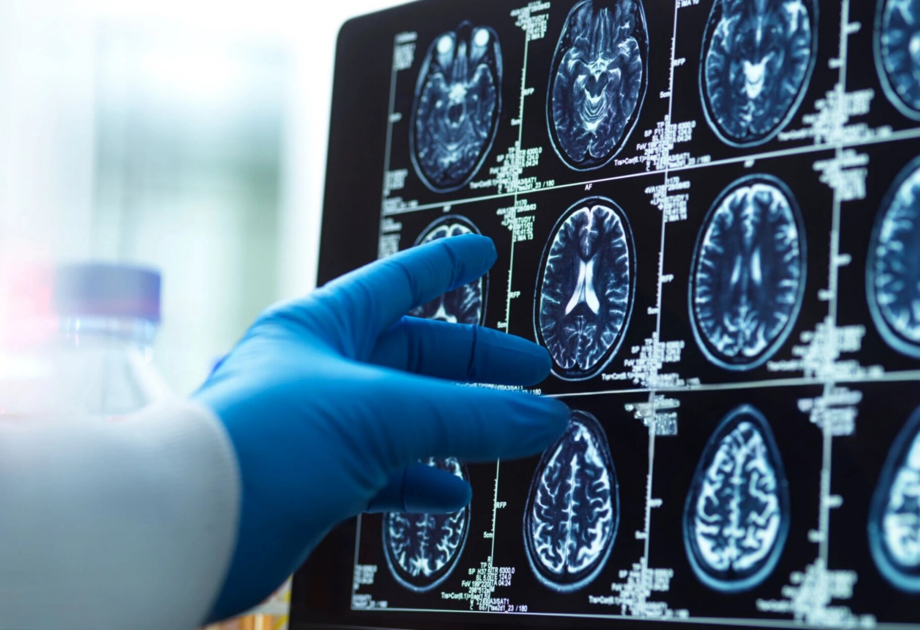
Clinicians use MRI scans of the brain as a tool to detect disease worsening in people with multiple sclerosis. (Photo by iStock)
- In analyzing brain MRIs of people with early relapsing-remitting multiple sclerosis, two markers were seen to accurately predict cognitive decline.
- These markers showed ongoing inflammation and a fivefold higher risk of symptom worsening independent of relapses or new lesions.
- Using these markers may help improve early risk assessment and guide personalized treatment strategies in RRMS, researchers say.
In analyzing brain MRIs on people with early relapsing-remitting multiple sclerosis (RRMS), two markers of ongoing inflammation may help identify those patients at a higher risk of cognitive decline despite having no relapses or new lesions, or signs of tissue damage, a study from Europe suggests.
The researchers say these markers showed strong accuracy in detecting individuals who are more likely to have worsening cognitive disability even without the relapses, or flares, that normally mark this type of multiple sclerosis.
According to the team, the study findings could help change the way clinicians address cognitive issues in RRMS.
“Previous research mainly conceptualized cognitive impairment as a static condition. … However, greater importance should be placed on the identification and prediction of cognitive decline over time,” the researchers wrote, noting that the two markers showed damage progression.
“These MRI markers merit further investigation as measures to improve early risk stratification and potentially enhance personalized therapeutic strategies aimed at preventing both physical and cognitive subtle disability accumulation,” the team wrote.
The study, “Paramagnetic Rim Lesions and Choroid Plexus Volume at Diagnosis Are Associated With Cognitive Progression Independent of Relapse and MRI Activity in Early Relapsing–Remitting Multiple Sclerosis,” was published in the journal Annals of Clinical and Translational Neurology.

MS is caused by inflammation in the brain and spinal cord that damages the myelin sheath, a protective layer around nerve fibers that helps them send electrical signals more efficiently. This can lead to a wide range of symptoms, including motor problems and cognitive impairment.
RRMS, its most common form, is marked by relapses — periods in which new symptoms appear or existing ones get worse — followed by recovery periods where symptoms ease or disappear.
Researchers looking for early markers of progression
However, disability worsening may occur independently of relapses or new MS lesions visible on MRI scans. Progression independent of relapse activity, or PIRA, “reflects chronic smoldering [low-grade] inflammation” that leads to neurodegeneration and irreversible damage, the researchers wrote.
Cognitive progression independent of relapses and MRI activity, known as cognitive PIRMA, is independent of physical disability and highly common in people with RRMS. As such, researchers are now looking for early markers that could identify people at higher risk of cognitive PIRMA.
“Among candidate MRI markers, paramagnetic rim lesions (PRLs) and choroid plexus (CP) enlargement have recently gained increasing attention,” the researchers wrote.
PRLs are areas at the edge of MRI lesions where inflammation remains active for long periods, reflecting ongoing immune activity that slowly continues to cause nerve damage and disability progression.
Choroid plexus is a network of cells and blood vessels that produces the fluid surrounding the brain and spinal cord. Its enlargement may indicate increased immune cell trafficking and sustained inflammatory activity, and it has been linked to PIRA.
Now, a research team led by scientists in Italy investigated whether PRLs and CP enlargement could help identify people with RRMS who are at greater risk of cognitive PIRMA.
Their study involved 87 people newly diagnosed with RRMS who underwent MRI scans at diagnosis and were followed up for an average of 6.2 years.

Markers indicate 5 times higher risk of cognitive decline
During that follow-up, nearly two-thirds (63%) of the patients showed meaningful cognitive decline, according to the team.
Importantly, for 80% of them, these changes occurred without relapses or MRI activity, the team noted.
The patients with cognitive PIRMA had a significantly higher median number of PRLs (1.5 vs. 1) and were significantly more likely to have at least one PRL (85% vs. 52%) than those whose cognitive function remained stable, the data showed. The cognitive PIRMA group also had a significantly higher choroid plexus volume (0.18% vs. 0.16%) compared with the cognitively stable group.
The overall burden of typical white matter lesions, which indicate damage to nerve fibers and myelin, did not differ between the two groups, the team noted.
Statistical analyses adjusted for potential influencing factors demonstrated that PRL presence and choroid plexus enlargement were each significantly linked to a five times higher risk of cognitive PIRMA.
Cognition should be considered and carefully monitored over time to reach a more adequate control on disease evolution and associated processes.
Each of these factors also allowed researchers to identify patients with cognitive PIRMA versus those without cognitive decline with about 80% accuracy, the researchers noted.
These findings indicate that “choroid plexus volume and PRLs may represent potential markers of silent cognitive progression,” according to the team.
“Cognitive progression could represent a window into smoldering processes that lead to chronic inflammatory processes,” the researchers wrote, noting that “cognition should be considered and carefully monitored over time to reach a more adequate control on disease evolution and associated processes.”
Among the study’s limitations, the team noted the small number of patients and limited follow-up time. The researchers emphasized that future studies should also include results regarding physical disability progression independent of relapses or MRI activity.




Tajuane Weaver
What is the actual meaning of Cognitive? And what should I look for as changes to my personal health since I have RRMS? Thanks in advance for your response