
New MRI technique may reveal signs of myelin repair in multiple sclerosis
Imaging changes were linked to improvements in disability scores over one year
Written by |

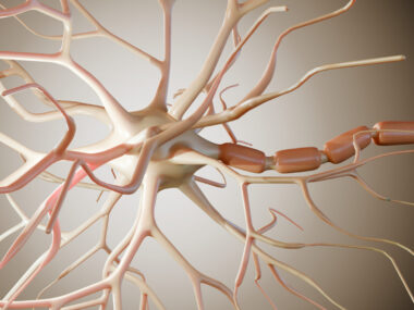
An illustration of the human brain. Researchers found that a specialized MRI technique may help detect signs of myelin repair in people with multiple sclerosis. (Photo from iStock)
- Multiple sclerosis is an inflammatory disorder that damages the myelin sheath surrounding nerve fibers in the brain and spinal cord.
- Conventional MRI scans cannot reliably measure myelin content, but a technique called APART-QSM can detect iron-sensitive and myelin-sensitive changes simultaneously.
- Increases in a myelin-sensitive MRI measurement were associated with improvements in disability scores, suggesting APART-QSM may help researchers monitor signs of myelin repair.
An MRI technique known as APART-QSM may help researchers track signs of repair to the myelin sheath, which is damaged in people with multiple sclerosis (MS), according to a study from China.
Researchers found that increases in a myelin-sensitive MRI measurement within MS lesions were significantly associated with improvements in disability scores. These changes may reflect myelin repair, a process called remyelination.
“APART–QSM may enable longitudinal monitoring of remyelination–related changes in MS,” the researchers wrote.
The study, “Monitoring Remyelination and Its Association With Disability Improvement in Multiple Sclerosis Using APART-QSM: A Longitudinal Cohort Study,” was published in the Journal of Magnetic Resonance Imaging.

New MRI technique may help detect signs of myelin repair
MS is an inflammatory disorder in which the immune system damages myelin, a fatty substance that surrounds nerve fibers and helps electrical signals travel quickly. On MRI scans, areas of damage appear as spots known as lesions.
MRI scans are an important tool for monitoring MS. However, while conventional MRI can detect areas of tissue damage, it cannot reliably measure how much myelin is present in brain tissue. More specialized techniques are needed to assess myelin content.
One such technique is APART-QSM, which stands for iterAtive magnetic suscePtibility sources sepARaTion quantitative susceptibility mapping. It uses computer-based analysis of MRI data to separate two types of magnetic susceptibility signals, called Chi-dia and Chi-para. The Greek letter chi is used to represent magnetic susceptibility.
Chi-dia is sensitive to myelin, while Chi-para is sensitive to iron. This distinction is important in MS because iron can accumulate in immune cells and contribute to inflammation and tissue damage.
“The ability of APART-QSM to separately characterize [Chi-para] and [Chi-dia] components allows iron-related pathology and myelin-sensitive changes to be assessed simultaneously within the same lesion,” the researchers wrote. “This distinction may be particularly valuable for monitoring remyelination, which is difficult to capture using conventional structural MRI alone.”
Researchers in China used APART-QSM to analyze MRI scans from 145 people with MS and 65 people without the disease, who served as controls. Of the MS patients, 101 underwent follow-up scans after a median of one year.
MRI changes linked to disability improvement in MS
Results showed that people with MS had significantly lower myelin-sensitive Chi-dia values and higher iron-sensitive Chi-para values than those without the disease in the putamen, a brain region involved in thinking and movement.
Within the MS group, white matter lesions showed significantly lower Chi-dia values and significantly higher Chi-para values than the surrounding normal-appearing white matter.
APART-QSM measurements also revealed changes over time. Chi-dia values increased within white matter lesions, potentially reflecting myelin repair, and greater increases were significantly associated with greater improvements in scores on a standard disability scale over about one year.
Greater increases in Chi-dia were also significantly associated with female sex and relapsing-remitting MS, data showed.
Chi-dia increases were also significantly greater in patients given the approved therapy Kesimpta (ofatumumab) than in untreated patients. The researchers cautioned that this finding should be interpreted carefully because patients in this observational study received individualized treatment regimens. They also noted that the MRI measurements were not directly validated against actual myelin and iron levels in tissue.
“APART-QSM revealed longitudinal increases in myelin-sensitive susceptibility within [white matter lesions] in patients with MS,” the researchers wrote. “These findings support further validation of susceptibility source separation MRI for individualized disease monitoring and evaluating repair-oriented therapeutic strategies in MS.”



Leave a comment
Fill in the required fields to post. Your email address will not be published.