
MRI maps reveal distinct brain network patterns in MS patients, study says
Technique could complement conventional MRI to help track disease activity
Written by |

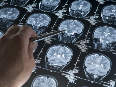
A doctor points at a patient's scan on a desktop computer monitor. (Photo from iStock)
- New MRI research shows that multiple sclerosis involves broader brain network reorganization rather than damage to isolated areas alone.
- Distinct brain network patterns correlate with specific clinical features, including disability, cognitive impairment, and disease activity.
- These individualized network maps could potentially improve multiple sclerosis diagnosis, tracking, and patient monitoring.
Patterns in how different regions of the brain are structurally organized may help distinguish people with multiple sclerosis (MS) from those without the disease and provide information about MS-related clinical features, a study in China found.
Using MRI scans, researchers created individualized maps of large brain networks and found widespread differences in people with MS compared with healthy people. Among those with MS, distinct brain network patterns were associated with disability, cognitive impairment, and disease activity.
The findings support the view of MS as a disease involving broader reorganization of brain networks, rather than damage to isolated areas alone, the researchers noted.
“Individualised network profiling may complement conventional MRI by improving characterisation of heterogeneity [variability] across disability, cognition, and disease activity, and by providing quantitative markers sensitive to subclinical [subtle] change in clinically stable patients,” the team wrote.
The study, “Brain network biomarkers for diagnosis and clinical stratification in multiple sclerosis: a longitudinal study integrating individualised structural covariance MRI with high-density EEG,” was published in eBioMedicine.

MRI plays central role in diagnosing, monitoring MS
MS is a chronic condition characterized by inflammation and damage to parts of the brain and spinal cord, leading to a range of symptoms that can affect movement, vision, sensation, and thinking.
MRI scans play a central role in establishing an MS diagnosis and monitoring disease progression, allowing doctors to identify lesions — areas of MS-related inflammation and damage. But these individual areas of damage don’t tell the whole story, according to the authors.
Research increasingly suggests that MS also alters larger networks of brain regions that work together to support complex functions such as movement, sensory processing, and thinking.
In this study, scientists in China investigated whether mapping these networks in individual patients could provide additional information about MS and its varied clinical manifestations.
They analyzed MRI scans from 635 adults across two independent groups. The main group included 163 people with MS and 248 healthy people recruited at Xuanwu Hospital in China. A second group from the UK Biobank — 117 people with MS and 107 healthy people — was used to see whether the findings could be reproduced in a separate group.
For each person, the researchers used structural MRI scans to build an individualized structural covariance network (iSCN). In simple terms, the technique examines gray matter — brain tissue rich in nerve cell bodies — across 170 brain regions to create a personalized map of how their structural characteristics relate to one another.

Widespread differences seen between MS patients, healthy people
The team examined two aspects of these maps: connectivity — the relationships between individual brain regions — and topology, which describes the network’s overall organization.
Compared with healthy people, those with MS showed widespread iSCN differences. These repeatedly involved the thalamus — a major relay center that helps information travel across the brain — other deep brain regions, and altered relationships in regions involved in visual processing.
Some relationships between brain regions were stronger in people with MS than in healthy people, while others were weaker, suggesting “that MS involves brain network reorganisation rather than a simple overall loss of connectivity,” the researchers wrote.
The extent of these abnormalities varied with patients’ clinical features. People with greater MS-related disability showed the most widespread changes, affecting overall network organization, individual brain regions, and structural relationships between them.
Cognitive impairment was associated with a more selective pattern of abnormalities involving the thalamus and cingulate regions, which relay and integrate information across the brain, as well as supplementary motor areas, which are involved in planning movement. Altered structural relationships involving the visual network were also seen.
[Findings suggest] that MS involves brain network reorganisation rather than a simple overall loss of connectivity.
Changes associated with disease activity involved a smaller set of brain regions that help process sensory information, emotions, and other complex functions.
Among 33 patients who met criteria for no evidence of disease activity — meaning no relapses, worsening disability, or new or enlarging MRI lesions — the MRI network analysis still detected subtle changes in individual brain regions over one year.
Although the analysis was small and “should be interpreted cautiously,” it suggests that subtle brain changes can still occur in a person who appears clinically stable, according to the authors.
“The iSCN approach may detect subclinical network evolution not captured by routine clinical or [MRI] assessments,” they wrote.
Artificial intelligence models were also used to test whether these network measures can distinguish people with MS from healthy controls and separate patients by clinical characteristics. Connectivity features were more informative for distinguishing MS from healthy controls, whereas network topology measures were generally more useful for separating MS patients by clinical features.
While more work is needed to validate the strategy, the “findings suggest that the iSCN approach can capture clinically meaningful brain reorganisation in MS and may help link routine MRI with future patient-level stratification,” the researchers concluded.



Umer Mirza
I have since 2011. I just had a relapse doctor think o am having heart attack but heart was fine. Than they find out I have scars in my medulla oblongata. That’s playing with my heart.
How can with get this MRI done and where.